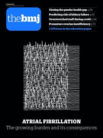
Temporal trends in lifetime risks of atrial fibrillation and its complications between 2000 and 2022

Antipsychotic use in people with dementia
Antipsychotic use in people with dementia is associated with elevated risks of a wide range of serious adverse outcomes
Antipsychotic use in people with dementia
Antipsychotic use in people with dementia is associated with elevated risks of a wide range of serious adverse outcomes

Child health: UK must regain “lost ground,” Kingdon urges
The outgoing RCPCH president is “ashamed” of the state of child health in the UK, she tells Gareth Iacobucci. But things can be turned around
Child health: UK must regain “lost ground,” Kingdon urges
The outgoing RCPCH president is “ashamed” of the state of child health in the UK, she tells Gareth Iacobucci. But things can be turned around

Closing the gender health gap: a £39bn boost to the economy, as well as lives
The UK has the 12th largest gender health gap in the world. Closing it will reap rewards for women and the country, reports Sarah Graham
Closing the gender health gap: a £39bn boost to the economy, as well as lives
The UK has the 12th largest gender health gap in the world. Closing it will reap rewards for women and the country, reports Sarah Graham
Trainees are removed from London surgery department after findings of bullying and poor supervision
Why hospital capacity is more complex than bed capacity
Nestlé accused of double standards over added sugar in baby products sold in poorer countries
Health under the spotlight in India’s 2024 election
Can a worker cooperative model of social care improve the patient experience and reduce pressure on GPs?
Mental health support for doctors—a vital yet fragile lifeline
Breaking free from the stigma of diabetes
Latest articles
News
Contaminated blood victims are still being let down by government, say campaigners
Community mental health services need support to improve patient satisfaction, says NHS leader
Medical device firms paid UK health organisations €37m in three years, study finds
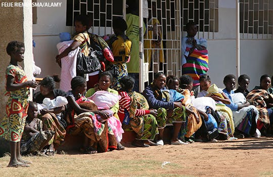
The world has made no progress in reducing preventable deaths in pregnancy and childbirth, says UN agency
GPs enter dispute with NHS England over contract imposition
Norway’s health minister resigns over thesis plagiarism allegations
Private GP provider is investigated for potential contract breaches
Psychiatrist who claimed £1000s for shifts he didn’t work is struck off
Smacking: Paediatricians call for ban in England and Northern Ireland
Research

TRIPOD+AI statement: updated guidance for reporting clinical prediction models that use regression or machine learning methods
Impact of large scale, multicomponent intervention to reduce proton pump inhibitor overuse
Esketamine after childbirth for mothers with prenatal depression
Glucagon-like peptide 1 receptor agonist use and risk of thyroid cancer
Quantifying possible bias in clinical and epidemiological studies with quantitative bias analysis: common approaches and limitations
Use of progestogens and the risk of intracranial meningioma
Delirium and incident dementia in hospital patients
Comment

Doctors should not be simply the ghost in the machine
Partha Kar: We need to level up all staff, not level down doctors
Helen Salisbury: With the NHS in crisis, why is Labour looking to private hospitals as the solution?
Health literacy matters
John Launer: Learning from appraisals
TRIPOD+AI: an updated reporting guideline for clinical prediction models
Why the fuss about physician associates?
Refocusing the WHO’s Model List of Essential Medicines on the needs of low and middle income countries
Predicting the outcomes of chronic kidney disease in older adults
Education

How to communicate about climate change with patients
Alternative routes into clinical research: a guide for early career doctors
Advances in management of heart failure
Non-acute heart failure management in primary care
Group A beta-haemolytic streptococcal infection in children
Secondary prevention of cardiovascular disease, including cholesterol targets: summary of updated NICE guidance
Effectiveness and safety of drugs for obesity
Dry eye disease management
Caring is the invisible piece of the stroke recovery puzzle
Stroke rehabilitation in adults: summary of updated NICE guidance
Workforce


BMJ Medicine


Practical implications of the ADNEX risk prediction model for diagnosis of ovarian cancer
Socioeconomic inequalities in risk of infection with SARS-CoV-2 delta and omicron variants in the UK, 2020-22
Preterm prelabour rupture of membranes before 23 weeks’ gestation
All cause and cause specific mortality associated with transition to daylight saving time in US
UK Jobs
Current issue
Most read
1.
Effect of exercise for depression: systematic review and network meta-analysis of randomised controlled trials
2.
Ultra-processed food exposure and adverse health outcomes: umbrella review of epidemiological meta-analyses
3.
Use of progestogens and the risk of intracranial meningioma: national case-control study
4.
Rosuvastatin versus atorvastatin treatment in adults with coronary artery disease: secondary analysis of the randomised LODESTAR trial
5.
Delirium and incident dementia in hospital patients in New South Wales, Australia: retrospective cohort study
6.


